■


As a library, NLM provides access to scientific literature. Inclusion in an NLM database does not imply endorsement of, or agreement with, the contents by NLM or the National Institutes of Health. Learn more about our disclaimer.
Sci Rep. 2021; 11: 14532.
Published online 2021 Jul 15. doi: 10.1038/s41598-021-93888-0
PMCID: PMC8282797
PMID: 34267258
Strictly regulated agonist-dependent activation of AMPA-R is the key characteristic of TAK-653 for robust synaptic responses and cognitive improvement
 1
1This article has been corrected. See Sci Rep. 2021 July 21; 11: 15255.
Associated Data
Abstract
Agonistic profiles of AMPA receptor (AMPA-R) potentiators may be associated with seizure risk and bell-shaped dose-response effects. Here, we report the pharmacological characteristics of a novel AMPA-R potentiator, TAK-653, which exhibits minimal agonistic properties. TAK-653 bound to the ligand binding domain of recombinant AMPA-R in a glutamate-dependent manner. TAK-653 strictly potentiated a glutamate-induced Ca2+ influx in hGluA1i-expressing CHO cells through structural interference at Ser743 in GluA1. In primary neurons, TAK-653 augmented AMPA-induced Ca2+ influx and AMPA-elicited currents via physiological AMPA-R with little agonistic effects. Interestingly, TAK-653 enhanced electrically evoked AMPA-R-mediated EPSPs more potently than AMPA (agonist) or LY451646 (AMPA-R potentiator with a prominent agonistic effect) in brain slices. Moreover, TAK-653 improved cognition for both working memory and recognition memory, while LY451646 did so only for recognition memory, and AMPA did not improve either. These data suggest that the facilitation of phasic AMPA-R activation by physiologically-released glutamate is the key to enhancing synaptic and cognitive functions, and nonselective activation of resting AMPA-Rs may negatively affect this process. Importantly, TAK-653 had a wide safety margin against convulsion; TAK-653 showed a 419-fold (plasma Cmax) and 1017-fold (AUC plasma) margin in rats. These findings provide insight into a therapeutically important aspect of AMPA-R potentiation.
Subject terms: Ligand-gated ion channels, Pharmacology, Drug discovery, Target validation
Introduction
Glutamate is the primary excitatory neurotransmitter throughout the central nervous system (CNS). Enhancement of glutamate-mediated transmission is critical for synaptic plasticity and for learning and memory1–3. Glutamatergic dysfunction has been implicated in various disorders including schizophrenia, Alzheimer’s disease (AD), attention-deficit/hyperactivity disorder (ADHD), autism and major depressive disorders; thus, potentiation of glutamatergic transmission could be a promising therapeutic strategy for psychiatric and neurological diseases4–6.
Multiple lines of evidence support activation of the alpha-amino-3-hydroxy-5-methyl-4-isoxazole-propionic acid receptor (AMPA-R) as a promising strategy for the treatment of CNS disorders7–11. However, AMPA-R agonists have a high seizure risk probably due to nonspecific activation of resting AMPA-R in the brain12–15, and hence are limited in their ability to secure a safety margin. As glutamate release in the brain is strictly regulated, AMPA-R potentiators (= AMPA-R positive allosteric modulators) which can promote physiological AMPA-R activation by glutamate have been considered as an alternative approach. However, AMPA-R potentiators such as LY451646 showed prominent agonistic effects (AMPA-R activation in the absence of agonist) in Ca2+ influx assays and whole-cell current recordings using primary hippocampal neurons16. Moreover, LY451646 had a narrow safety margin against seizures and a narrow bell-shaped dose-response for cognitive improvement in rats and monkeys16. A bell-shaped dose-response was also seen in various other pharmacological assays of the AMPA-R potentiators LY451646 and S1898617–19. The bell-shaped dose-response of AMPA-R potentiators may be due to desensitization of AMPA-R; however, this hypothesis is inconsistent with their seizure induction at higher doses. Thus, further characterization of the mechanisms of action underlying the bell-shaped dose-response effect of AMPA-R potentiators is needed.
We recently reported that structural interference at Ser750 in AMPA-R GluA2o (corresponding to Ser743 in GluA1i) was key to lowering the agonistic effect of AMPA-R potentiators containing a dihydropyridothiadiazine 2,2-dioxides skeleton16. To discover novel AMPA-R potentiators with better pharmacological profiles, we established an original screening strategy consisting of 1) binding assays with or without glutamate using [3H]-HBT1 and purified recombinant ligand-binding domain (LBD) of AMPA-R, 2) X-ray crystallography, 3) Ca2+ influx assays using wild-type hGluA1i or hGluA1i with a mutation at Ser743 in CHO cells, 4) Ca2+ influx assay in rat primary hippocampal neurons, and 5) whole-cell patch clamp recording using rat primary hippocampal neurons. By using the screening strategy, we designed and searched for AMPA-R potentiators with lower agonistic effects and discovered TAK-137. TAK-137 bound to AMPA-R in a glutamate-dependent manner and showed a lower agonistic effect than LY451646 in both Ca2+ influx assay and whole-cell patch clamp recording in rat primary hippocampal neurons. Studies in rats found potent cognitive improvement and a wider safety margin against seizures with TAK-137 as compared with LY451646. However, TAK-137 still maintained a weak agonistic effect16,20.
AMPA-R potentiators with lower agonistic effects than TAK-137 have not been explored yet; such a compound may have a wider safety margin against seizures or may lose beneficial effects, including cognitive improvement. Thus, we decided to discover and characterize a novel AMPA-R potentiator with lower agonistic effects than TAK-137. Through our original screening strategy, we discovered TAK-653, 9-[4-(Cyclohexyloxy)phenyl]-7-methyl-3,4-dihydropyrazino[2,1-c][1,2,4]thiadiazine 2,2-dioxide, which had virtually no agonistic effects in primary hippocampal neurons. Here, we describe the pharmacological characteristics of TAK-653. Our data suggest that the facilitation of phasic AMPA-R activation by physiologically-released endogenous agonist (glutamate) is the key to enhance synaptic and cognitive functions, and nonselective activation of resting AMPA-Rs by agonistic effects may negatively affect both synaptic and cognitive functions, resulting in bell-shaped dose-response effects.
Results
TAK-653 bound to the LBD of purified recombinant AMPA-R proteins and induced Ca2+ influx in hGluA1i CHO cells in a glutamate-dependent manner
We previously reported that structural interference at Ser750 in the channel-closed state of GluA2o LBD might be involved in the molecular mechanisms underlying the lower agonistic effect of AMPA-R potentiators with dihydropyridothiadiazine 2,2-dioxide derivatives and discovered TAK-137, 9-(4-phenoxyphenyl)-3,4-dihydropyrido[2,1-c][1,2,4]thiadiazine 2,2-dioxide (Fig. 1A)16. Based on the structure and agonistic-effect relationship studies on this novel chemical series, we designed TAK-653, a dihydropyrazinothiadiazine 2,2-dioxide derivative with cyclohexyl group as bulky terminal substituents to induce steric repulsion at Ser750 in GluA2o; the structural bulkiness of cyclohexyl group is higher than that of the terminal phenyl group of TAK-137, suggesting that TAK-653 may exhibit a lower agonistic effect. TAK-653 bound to the intradimer interface formed by the ligand binding core (Fig. 1B). The 3D overlay analysis showed that the peripheral cyclohexyl rings of TAK-653 caused steric interference at Ser750 in the channel-closed state of GluA2o LBD (Toyofuku et al., in preparation). Binding affinity of TAK-653 to the GluA2o LBD was measured by a scintillation proximity assay (SPA) using [3H]-HBT1, a radio-labeled LBD-binding AMPA-R potentiator, and a His-tagged GluA2o LBD protein (His-LBD)21. TAK-653 inhibited binding between [3H]-HBT1 and His-LBD with an IC50 value of 0.26 μM (Fig. 1C). Binding of [3H]-TAK-653 to His-LBD was also measured by SPA. Binding between [3H]-TAK-653 and His-LBD was robustly increased in a glutamate-dependent manner, whereas binding of [3H]-TAK-653 was not detected when another His-tagged protein such as macrophage migration inhibitory factor (MIF) was used as a control (Fig. 1D). TAK-653 did not inhibit the specific binding of [3H]-AMPA to His-LBD, but rather mildly increased [3H]-AMPA binding with EC50 and Emax values of 1.5 ± 0.2 μM and 19.9 ± 3.4%, respectively (Fig. 1E), suggesting that TAK-653 has no binding affinity for the agonist binding site of AMPA-R. These results suggest that TAK-653 selectively binds to the LBD of AMPA-R in a glutamate-dependent manner due to structural interference at Ser750 (GluA2o LBD) in the channel-closed state.

Selective binding of TAK-653 to AMPA-R in a glutamate-dependent manner. (A) Chemical structure of TAK-653 and TAK-137. (B) The GluA2 (flop)/glutamate/TAK-653 complex crystallized as a dimer. (C) Displacement studies with TAK-653 using the SPA assay with [3H]-HBT1 and His-LBD. Data are represented as mean ± SD (n = 4). (D) Effects of glutamate on the binding of [3H]-TAK-653 to His-LBD. Data are represented as relative value ± SD in counts per minute (CPM) (n = 3). (E) Effect of TAK-653 on the binding of [3H]-AMPA to His-LBD. Data are represented as mean ± SEM (n = 4). (F) Effects of TAK-653 on Ca2+ influx in GluA1i CHO cells in the presence (open circle) and absence (open square) of 3 mM glutamate. Data are represented as mean ± SD (n = 3). (G) Effects of TAK-653 on Ca2+ influx in CHO cells expressing GluA1i WT or GluA1i S743A in the absence or presence of glutamate (0.3 μM, 1 μM and 3 mM). S743 in GluA1i LBD corresponds to S750 in GluA2o LBD. Data are represented as mean ± SD (n = 3). (H) Effects of TAK-653 on Ca2+ influx in hGluA1i CHO cells (closed circle) and rGluA1i CHO cells (open circle) in the presence of 3 mM glutamate. Data are represented as mean ± SD (n = 3).
In the functional assays, TAK-653 robustly increased Ca2+ influx only in the presence of glutamate (3 mM) in hGluA1i CHO cells; the EC50 was 3.3 μM (Fig. 1F). Ser750 in GluA2o LBD corresponds to Ser743 in GluA1i LBD, thus the introduction of an S743A mutation into GluA1i was expected to reduce the steric interference with the peripheral cyclohexyl rings of TAK-653 and to facilitate the binding of TAK-653 to GluA1i especially when glutamate concentration is low. In fact, the maximum responses of TAK-653 in CHO cells expressing S743A GluA1i were higher than those in CHO cells expressing wild-type GluA1i in a Ca2+ influx assay at each concentration of glutamate tested (Fig. 1G). Next, to improve the prediction of efficacious concentration for use in humans, we assessed potential of TAK-653 in human and rat GluA1i. In a Ca2+ influx assay using human and rat GluA1i-expressing CHO cells, TAK-653 did not show species differences; the fold difference in the EC50 value between rat and human receptors was 1.1 (Fig. 1H). TAK-653 did not show prominent subunit selectivity for homomeric AMPA-R in a Ca2+ influx assay using CHO cells expressing GluA1-4i and TARP γ-2 or GluA1-4o and TARP γ-2 (Table S1). TAK-653 at 10 μM was highly selective against 97 targets (Ricerca, Taipei, Taiwan); TAK-653 only inhibited lipoxygenase 5-LO enzyme activity with an IC50 value of 5.9 μM (Table S2). These observations further suggest that TAK-653 selectively binds to AMPA-R in a glutamate-dependent manner due to structural interference at Ser750 in GluA2o LBD (S743A in GluA1i LBD).
TAK-653 induced Ca2+ influx, whole-cell currents, and brain-derived neurotrophic factor (BDNF) production in an AMPA-dependent manner in primary hippocampal neurons
In our previous studies, recombinant AMPA-R on hGluA1i CHO cells was less sensitive than physiological AMPA-R on primary neurons in the detection of agonistic effects of AMPA-R potentiators16,21. Thus, we next characterized TAK-653 using rat primary hippocampal neurons. A single application of AMPA dose-dependently increased intracellular Ca2+ levels (Fig. 2A). Thus, our experimental conditions are suitable for the characterization of AMPA-R potentiators using cultured primary neurons. The response of 5 μM AMPA plus 10 μM HBT1 was defined as 100% in the Ca2+ influx assay using rat primary hippocampal neurons21. TAK-653 robustly increased Ca2+ influx only in the presence of AMPA (5 μM) with an EC50 of 0.93 μM (Fig. 2B). TAK-653, TAK-137, and LY451646 at 30 μM achieved the maximal Ca2+ increase in the presence of agonist (AMPA), thus the response at 30 μM was used to compare their agonistic effects. The Ca2+ increase produced by TAK-653, TAK-137, and LY451646 at 30 μM was 4.8%, 7.6%, and 88%, respectively (Table (Table11)16.

Effect of TAK-653 on Ca2+ influx and AMPA-R currents in rat primary hippocampal neurons. (A) Effect of AMPA on Ca2+ influx in primary hippocampal neurons. (B) Effect of TAK-653 on Ca2+ influx in primary hippocampal neurons. TAK-653 was applied in the presence or absence of 5 μM AMPA. Data are represented as mean ± SD (n = 3). (C) Effects of AMPA on AMPA-R-mediated currents using primary hippocampal neurons. (D, E) Effects of TAK-653 on AMPA-R-mediated currents using primary hippocampal neurons. The responses to TAK-653 were measured in the presence (D) or absence (E) of 1 μM AMPA. AMPA-R potentiator was applied 20 s prior to 10-s AMPA stimulus. Without AMPA, the maximum current was measured during 60-s application. Data are represented as mean ± SEM (n = 6).
Table 1
Effects on intracellular Ca2+ level and AMPA-R-mediated currents in the presence or absence of AMPA in rat primary hippocampal neurons.
| Intracellular Ca2+ level * | AMPA-R currents # | |||
|---|---|---|---|---|
| Potentiation EC50 (μM) | Agonistic effect @30 μM (%) | Potentiation EC50 (μM) | Agonistic effect @30 μM (%) | |
| TAK-653 | 0.93 | 4.8 | 4.4 | 1.7 |
| TAK-137 | 0.42 | 7.6 | 1.4 | 6.4 |
| LY451646 | 0.78 | 88 | 1.9 | 39 |
*% of 5 μM AMPA + 10 μM HBT1 response.
# % of AMPA (100 μM)-induced steady-state response.
EC50 value was calculated from dose-response curve in the presence of 5 μM AMPA for intracellular Ca2+ levels or 1 μM AMPA for AMPA-R currents using a nonlinear regression.
In whole-cell patch clamp recordings using rat primary hippocampal neurons, a single application of AMPA dose-dependently increased AMPA-R-mediated currents with an EC50 value of 45 μM (Fig. 2C). Thus, AMPA at a low dose of 1 μM was used to characterize the AMPA-R potentiation activity of TAK-653. The response of TAK-653 with AMPA (1 μM) was normalized against currents induced by 1 μM AMPA. TAK-653 dose-dependently augmented AMPA (1 μM)-elicited currents with an EC50 value of 4.4 μM (Fig. 2D). Next, we assessed agonistic effect of TAK-653. In the absence of agonist, the response of TAK-653 was normalized against currents induced by 100 μM AMPA. As a result, TAK-653 even at 30 μM produced 1.7% of the AMPA (100 μM)-elicited currents, while TAK-137 and LY451646 showed 6.4% and 39%, respectively, response at 30 μM (Fig. 2E and Table Table11)16,20. Thus, the agonistic effects of TAK-653 were lower than those of TAK-137 and LY451646 in rat primary hippocampal neurons.
Activation of AMPA-R increased BDNF mRNA levels in the mouse hippocampus22,23. We examined the effect of TAK-653 on BDNF production in rat primary hippocampal neurons. TAK-653 robustly increased BDNF protein levels in the presence of a low concentration of AMPA (1 μM) (Fig. 3A). TAK-653 alone slightly increased BDNF protein levels at 1 μM. Next, we investigated the effect of TAK-653 on BDNF mRNA levels in the mouse hippocampus. TAK-653 at 3 and 10 mg/kg, p.o. significantly increased BDNF mRNA levels in the AMPA (3.5 mg/kg, i.v.)-treated mice, while TAK-653 alone did not increase BDNF mRNA levels under these experimental conditions (Fig. 3B). These results demonstrate that TAK-653 stimulates BDNF production through agonist-dependent AMPA-R activation in both in vitro and in vivo.

Effects of TAK-653 on BDNF expression in vitro and in vivo. (A) Effects of TAK-653 on BDNF protein levels in rat primary hippocampal neurons. Cells were treated with AMPA (0 or 1 μM) and TAK-653 (0.01, 0.1, 1 μM) for 24 h and then were collected using lysis buffer. Cells in control group were treated with 1 μM AMPA and DMSO. Values were expressed as pg per mL. Data are represented as mean ± SD (n = 3). Statistical significance was determined by a two-tailed Williams’ test with significance set at #P ≤ 0.05 (versus control group; two-tailed Williams’ test). (B) Effects of TAK-653 on BDNF mRNA in hippocampus in AMPA (3.5 mg/kg, i.v.)-treated mice. TAK-653 (3 and 10 mg/kg, p.o.) was administered to mice 1 h before the administration of AMPA (3.5 mg/kg, i.v.) (left) or vehicle (right). Tissues were isolated 3 h after AMPA administration. Data were presented as the mean ± SEM (n = 23–24). #P ≤ 0.05 (versus vehicle-treated group; two-tailed Williams’ test).
TAK-653, but not LY451646 or AMPA, improved cognitive functions in multiple domains in rats and working memory in monkeys
The effect of TAK-653 on visual learning and memory was assessed using the novel object recognition (NOR) test in rats. TAK-653 at 0.03, 0.1 and 0.3 mg/kg, p.o. significantly improved the novelty discrimination index (NDI) (Fig. 4A). TAK-653 may enhance visual learning and memory at ≥ 0.03 mg/kg, p.o. in normal rats. In our previous study in rats, LY451646 at ≥ 1 mg/kg, p.o. improved visual learning and memory in the NOR test, although it induced seizure at 10 and 30 mg/kg, p.o.16. AMPA induced seizure at 30 mg/kg, i.p. and abnormal behavior such as head turning at 10 mg/kg, i.p. in rats, thus we assessed its effects on cognitive function at 3 mg/kg, i.p. and less. Surprisingly, AMPA at 0.3 to 3 mg/kg, i.p. did not improve cognitive performance in the NOR test (Fig. 4B).

Effect of TAK-653, LY451646, and AMPA on cognitive functions in multiple domains. (A) Effect of TAK-653 on visual learning and memory in NOR test using naïve rats. TAK-653 (0.01 and 0.03 mg/kg, p.o., 0.03 and 0.1 mg/kg, p.o., and 0.1 and 0.3 mg/kg, p.o.) was administered to rats 2 h prior to the acquisition and the retention trials. (B) Effect of AMPA on visual learning and memory in NOR test using naïve rats. AMPA (0.3, 1 and 3 mg/kg, i.p.) was administered 0.5 h prior to the acquisition and the retention trials. NDI data were presented as the mean ± SEM (n = 10). #P ≤ 0.05 (versus vehicle-treated group; two-tailed Williams’ test). (C–E) Effect of TAK-653 (C), LY451646 (D) or AMPA (E) on working memory in RAM test using rats. At 1.5 h, 1 h or 0 h before administration of vehicle or MK-801 (0.08 mg/kg, s.c.), TAK-653, LY451646 or AMPA, respectively, was administered to rats. Thirty minutes after dosing of MK-801, rats were placed on the maze, and then the entry into the arm was recorded. The mean errors were indicated as the mean ± SEM (n = 6–18). ***P ≤ 0.001 (versus vehicle-vehicle group; Welch’s test); #P ≤ 0.05 (versus vehicle-MK-801 group; two-tailed Shirley-Williams’ test). (F) Effect of TAK-653 on working memory in DMTS test using monkeys. TAK-653 at 0.06 mg/kg was orally administered to monkeys at 6 h prior to DMTS testing. Each plot at 0, 4, 8 or 16 s interval were presented as the mean ± SEM within 96 trials per session (n = 3) (left). TAK-653 significantly ameliorated DMTS accuracy at 16 s delay interval (right). *P ≤ 0.05 (versus vehicle group; paired t test). (G) Effect of TAK-653 on sociability deficits in the social approach-avoidance test in poly-I:C mouse. TAK-653 at 0.3 mg/kg was orally administered to mice at 1 h prior to test session. Sniffing index data were presented as the mean ± SEM (n = 7). *P ≤ 0.05 (versus control mice group; Student’s t test); #P ≤ 0.05 (versus control mice group; Student’s t test).
The effect of TAK-653 on MK-801-induced working memory deficit in rats was evaluated using the radial arm maze (RAM) task. MK-801 (0.08 mg/kg, s.c.) disrupted the performance of well-trained rats and TAK-653 at 0.1, 0.3, 3 and 10 mg/kg, p.o. significantly ameliorated the MK-801-induced deficits (Fig. 4C). Thus, TAK-653 may enhance working memory performance over a broad dose range in hypoglutamatergic conditions. By contrast, neither LY451646 at 0.03 to 1 mg/kg, p.o. nor AMPA at 0.1 to 3 mg/kg, i.p. improve working memory in the RAM test (Fig. 4D,E).
We also characterized another AMPA-R potentiator, PF-04958242. PF-04958242 showed significant agonistic effects; PF-04958242 potently produced a Ca2+ increase in the absence of AMPA in rat primary hippocampal neurons (Fig. S1A). Similar to LY451646, PF-04958242 improved visual learning and memory at ≥ 0.1 mg/kg, p.o. in the NOR test, while PF-04958242 did not improve working memory in the RAM test (Fig. S1B and S1C).
The effect of TAK-653 on working memory was also assessed using the delayed match-to-sample (DMTS) paradigm in monkeys. Cmax at 0.06 mg/kg p.o. in fasted monkeys corresponds to that at 0.1 mg/kg p.o. in rats, thus 0.06 mg/kg, p.o. of TAK-653 was used in the monkey study. TAK-653 at 0.06 mg/kg, p.o. significantly increased DMTS accuracy at a 16-s delay interval (Fig. 4F). The significant improvement in accuracy returned to the vehicle level at 48 h later, reflecting that the favorable effect was not attributable to a training effect. The beneficial effect of TAK-653 on task accuracy maintained 24 h after administration probably due to its Tmax value (8.0 ± 0.0 h) and t1/2 value (9.4 ± 3.8 h) at 0.03 mg/kg, p.o. in monkeys. TAK-653 may also improve working memory in monkeys at similar plasma concentrations as observed in rats for improving visual learning, recognition memory and working memory.
We investigated the effects of TAK-653 on attention in the rat 5-choice serial reaction time task (5CSRTT). Rats with poor performance could be a useful model of ADHD24. Thus, sub-population analyses with a median split of the population into high and poor performing animals, were also performed based on their correct responses under vehicle treatment for each treatment block25. TAK-653 (0.3 mg/kg, p.o.) administration at 2 h prior to the trial showed no significant improvement in the whole population, but median split analysis (median value, 51) revealed that TAK-653 significantly increased correct responses and decreased omissions in the poor performing rats (Fig. S2). TAK-653 did not affect the premature responses. Thus, TAK-653 may enhance sustained attention in the poor performing rats.
The effect of TAK-653 on sociability deficits was evaluated using the social approach-avoidance test in the poly-I:C mouse, a developmental immune activation animal model of schizophrenia26,27. The sniffing index was significantly decreased in vehicle-treated poly-I:C mice compared with vehicle-treated control mice. TAK-653 at 0.3 mg/kg, p.o. significantly improved the sniffing index (Fig. 4G). TAK-653 may ameliorate abnormal social interaction.
TAK-653 had a low risk of receptor desensitization or sensitization in vivo
Down-regulation of AMPA-Rs or sensitization of the AMPA-R system following chronic simulation are a concern with AMPA-R activators28,29. TAK-653 at 0.3 mg/kg, p.o. improved social interaction in mice (Fig. 4G) and in vivo plasma exposure level at 0.1 mg/kg, p.o. were similar between rats and mice (Table S3), thus we assessed AMPA-R function after 14 days of TAK-653 administration at 0.3 mg/kg p.o. in mice. AMPA-R activation is known to induce the expression of BDNF and growth arrest and DNA-damage-inducible beta (Gadd45b) mRNA30. Pre-administration of TAK-653 at 0.3 mg/kg, p.o. for 14 days did not affect AMPA-induced BDNF and Gadd45b mRNA expression in the mouse hippocampus (Fig. 5A,B), suggesting a lower risk of receptor desensitization or sensitization after repetitive dosing.

Effects of repeated treatment of TAK-653 on AMPA-induced BDNF (A) or Gadd45b (B) mRNA expression in mouse hippocampus. Vehicle or TAK-653 (0.3 mg/kg, p.o.) for 14 days were administered to mice. On the day 14, vehicle or AMPA (1.25, 2.5, 5 or 10 mg/kg, i.v.) was administered 1 h after vehicle (left) or TAK-653 (right). Tissues were isolated 3 h after AMPA administration. Data were presented as the mean ± SEM (n = 23–24). #P ≤ 0.05 (versus vehicle-treated group; two-tailed Shirley–Williams test).
TAK-653 enhanced AMPA-R-mediated synaptic responses more potently than AMPA or LY451646 in prefrontal cortex (PFC) slices
Desensitization of AMPA-R by agonistic effects may be a cause for reduced (or lack of) efficacy or extremely narrow bell-shaped dose-responses in cognitive improvement mediated by AMPA or LY451646; however, this hypothesis is inconsistent with their seizure risk at higher doses. To understand the underlying mechanism of action, we examined AMPA-mediated synaptic responses. In the presence of bicuculline (20 μM), CGP52422 (10 μM), and APV (50 μM) to block GABAA, GABAB and NMDA receptors, respectively, electrical stimulation of layer I elicited the AMPA-R-mediated polysynaptic EPSPs of layer V pyramidal neurons into burst firing while the somatic membrane was maintained at approximately − 65 mV with constant current (Fig. 6A). Bath application of 0.3 to 30 μM TAK-653 for 10 min enhanced the suprathreshold polysynaptic EPSPs with a significant increase in evoked spikes (Fig. 6B) and EPSP duration (Fig. 6C) in a concentration-dependent manner. The enhancing effects of TAK-653 were maintained for at least 10 to 20 min after elimination. By blocking the synaptic responses of AMPA-R with NBQX (10 μM), the effect of TAK-653 completely disappeared (Fig. 6A). Similar to TAK-653, application of TAK-137 at 3 μM also robustly enhanced polysynaptic EPSP duration and evoked spikes (Fig. S3). Compared with TAK-653 and TAK-137, LY451646 at 0.3 to 30 μM induced moderate potentiation of suprathreshold polysynaptic EPSPs with a smaller number of spikes (Fig. 6D) and EPSP duration (Fig. 6E). Following a switch from LY451646 (30 μM) to TAK-653 (10 μM) (Fig. 6F), a clear increase in spike number (Fig. 6G) and EPSP duration (Fig. 6H) were observed, indicating no pronounced desensitization of AMPA-R by LY451646. Both TAK-653 and LY451646 did not increase the membrane potentials of recorded neurons, while AMPA (0.3 μM) increased the membrane potentials. Under these conditions, AMPA reduced polysynaptic EPSP duration and evoked spike number when the membrane was held at − 65 mV with a current injection (Fig. 6I). Thus, TAK-653 might augment the AMPA-R-mediated polysynaptic network interactions more robustly than LY451646 or AMPA.

Effect of TAK-653, LY451646, and AMPA on AMPA-R-mediated EPSPs in prefrontal cortical slice. (A) In the presence of bicuculline (20 μM), CGP52422 (10 μM), and APV (50 μM) to block GABAA and B and NMDA receptors, respectively, single stimulation of glutamatergic afferents to PFC pyramidal neurons evoked polysynaptic EPSPs to induce action potential spiking. Addition of 10 μM TAK-653 for 10 min enhanced the suprathreshold response, resulting in the prolongation of action potentials trains. This response was abolished by the AMPA receptor antagonist NBQX (10 μM). (B, C) Effects of TAK-653 on the number of spikes (B) and duration (C) of AMPA-R-mediated EPSPs. Data were represented as mean ± SEM (n = 6–10). (D, E) Effects of LY451646 on the number of spikes (D) and duration (E) of AMPA-R-mediated EPSPs. (F) Addition of 30 μM LY451646 for 10 min enhanced the suprathreshold response. Perfusion changes from LY451646 to TAK653 caused further augmentation of spike number. (G, H) Effects of TAK-653 after removal of LY451646 on the number of spikes (G) and duration (H) of AMPA-R-mediated EPSPs. Data were represented as mean ± SEM (n = 6–9). Statistical significance between LY451646 and TAK-653 was determined by a paired t test with significance set at #P ≤ 0.05. (I) Application of 0.3 μM AMPA for 5 min diminished the suprathreshold response (n = 6).
TAK-653 had a wider safety margin against seizures than LY451646 in rats
Cognitive improvement by TAK-137 in a variety of paradigms was observed at a similar dosage (around 0.1 mg/kg) in rats16,31. On the other hand, LY451646 showed dose-dependent efficacy only in the NOR test. Thus, we decided to calculate the exposure safety margins of AMPA-R potentiators based on the cognitive improvement in the NOR test and the absence of signs of seizures. TAK-653 elicited chronic and tonic convulsions in 1 animal at 4 h after dosing at 100 mg/kg, p.o. (Table S4). The exposure margins of TAK-653, calculated using the area under the plasma drug concentration–time curve (AUCplasma) values and plasma Cmax values, were 1017-fold (AUCplasma) and 419-fold (plasma Cmax), respectively (Table (Table22 and S5). Exposure margins of TAK-137 and LY451646 by this protocol were 122- and 4.0-fold (AUCplasma), respectively, and 42- and 3.4-fold (plasma Cmax), respectively. Thus, TAK-653 may have a wider exposure margin than LY451646 in rats.
Table 2
Exposure margin against seizure after acute treatment in rats (plasma Cmax and AUCplasma).
| TAK-653 | TAK-137 | LY451646 | |
|---|---|---|---|
| Margin based on plasma Cmax (fold) | 419 | 42 | 3.4 |
| Margin based on AUCplasma (fold) | 1017 | 122 | 4.0 |
Discussion
In neuropsychiatric disorders, functional enhancement of AMPA-R has the potential to improve cognitive deficits in several cognitive domains including executive function, attention, and working memory32. Enhancement of AMPA-R-mediated neurotransmission may also lead to rapid antidepressant action9,33–35. Thus, AMPA-R potentiators could be promising therapeutic drugs for multiple CNS disorders. However, seizure liability and narrow bell-shaped dose-responses might have restricted the development of AMPA-R potentiators as therapeutic drugs.
We hypothesized that the agonistic effects of some AMPA-R potentiators are associated with their seizure risks and bell-shaped dose-response effects. In fact, TAK-137, an AMPA-R potentiator with lower agonistic effects than LY451646, showed lower risks of seizure and bell-shaped dose-response16. In this study, we asked whether an AMPA-R potentiator with lower agonistic effects than TAK-137 (i.e. an AMPA-R potentiator with virtually no agonistic effect) may have lower seizure risks or if the beneficial effect is lost. To answer this question, we discovered a novel AMPA-R potentiator TAK-653 with extremely lower agonistic effects. [3H]-TAK-653 bound to the intradimer interface formed by the ligand-binding core of AMPA-R subunits in a glutamate-dependent manner. This intradimer interface is known to undergo conformational changes upon glutamate binding36,37. Co-crystallization studies suggested that TAK-653 exhibited glutamate-dependent structural interference at Ser750, located in the intradimer interface of GluA2o with larger steric repulsion in the absence of glutamate. In fact, the introduction of a mutation to Ser743 (S743A) in GluA1i (corresponding to Ser750 in GluA2o) to lower the steric interaction between AMPA-R and TAK-653 reduced the glutamate threshold for AMPA-R activation by TAK-653. Thus, like TAK-137, steric interaction at Ser743 in the channel-closed state may be related to the lower agonistic effects of TAK-65316. Both TAK-137 and TAK-653 had similar potential in the augmentation of agonist-elicited AMPA-R function, while TAK-653 had a lower agonistic effect than TAK-137 on both Ca2+ influx and whole-cell currents using primary neurons.
Quite interestingly, like TAK-137, TAK-653 produced a potent cognitive improvement in a wide range of cognitive domains. Neither LY451646 nor AMPA, however, improved working memory in the RAM test and AMPA did not improve recognition memory in the NOR test in rats. The effects of LY451646 in the RAM test were evaluated at around 1 mg/kg, p.o., a dose at which LY451646 produced efficacy in the NOR test. Therefore, brain exposure of LY451646 under the conditions for the RAM test was high enough to explore its therapeutic potential for working memory. The effects of AMPA were investigated at a broader range of doses from 0.3 to 3 mg/kg, i.p. because AMPA produced abnormal behavior at 10 mg/kg, i.p. Note that all three compounds induced convulsions at higher exposure, thus AMPA-R activation by all of them was clear. Therefore, facilitation of physiologically active AMPA-Rs, but not increasing the number of active AMPA-Rs by stimulating resting receptors, is key to producing potent cognitive improvements through AMPA-R activation. In line with this hypothesis, PF-04958242, with prominent agonistic activity for AMPA-R on rat primary hippocampal neurons, improved visual learning and memory but it did not improve working memory.
In isolated primary neurons, AMPA dose-dependently increased intracellular Ca2+ levels and AMPA-R-mediated currents (Fig. 2). Under these conditions, TAK-653 and LY451646 showed similar activity in the potentiation of AMPA-elicited Ca2+ increases and TAK-653 was 2.3 times less potent than LY451646 in the potentiation of AMPA-elicited currents (Table (Table11 and fig. S4). To our surprise, TAK-653 produced more robust potentiation of the AMPA-R component of the synaptic responses, especially the spike number, compared with LY451646 in the PFC neuronal network (Fig. 6). TAK-653 could enhance polysynaptic EPSPs after LY451646 exposure, thus it is unlikely that LY451646 reduced the number of AMPA-Rs by rapid desensitization during the 10 min for slice preparation. Furthermore, AMPA (0.3 μM) reduced polysynaptic EPSP duration and evoked spikes. Most neurons transform thousands of synaptic inputs into specific patterns of action potential output38,39. Nonspecific stimulation of resting AMPA-R by agonistic effects could activate multiple neural circuits simultaneously. The resulting continuous and repetitive synaptic inputs on single neurons connected through their multiple pathways may cause sublinear summation, such as through the activation of postsynaptic conductance40,41, with the reduced signal-to-noise ratio of an evoked response. TAK-653 might remarkably increase firing rate via strictly regulated agonist-dependent activation of AMPA-R, which may lead to potent cognitive improvement (Table S7). In line with this observation, similar to TAK-653, TAK-137, which improves cognitive functions in multiple paradigms, also robustly potentiated the AMPA-R component of the synaptic responses.
As previously reported, TAK-137 has little agonistic activity. However, compared with TAK-137, TAK-653 may have even lower agonistic activity. TAK-653 has a wider exposure margin than TAK-137 between cognitive improvement and seizure in rats, thus, this small difference in the agonistic effects of TAK-653 and TAK-137 in vitro likely related to their seizure susceptibility at higher dose ranges. Besides seizure liability, a bell-shaped dose-response might have limited the development of AMPA-R potentiators as drugs42,43. Given the heterogeneity of human metabolic profiles that may be associated with larger variations in pharmacokinetic profiles of drugs, bell-shaped dose-responses may be a significant disadvantage even with deliberate design of dose selection44. TAK-653 may overcome these issues because it showed cognitive improvements at a wider dose range and had a low risk of receptor desensitization or sensitization after 14 days of repetitive dosing.
Multiple findings have suggested that ketamine can produce antidepressant activity through a rapid disinhibition of pyramidal neurons and subsequent glutamate burst, followed by postsynaptic AMPA-R activation45. Based on the enhancement of AMPA-R-mediated EPSPs, TAK-653 could robustly enhance the activity of postsynaptic AMPA-Rs. These data further support the potential antidepressant action of TAK-653, although detailed preclinical studies to assess antidepressant effects of TAK-653 are needed.
In summary, we found that agonistic effects may significantly impair the function of AMPA-R potentiators in synaptic transmission and interfere with cognitive improvement. In fact, TAK-653, an AMPA-R potentiator with minimal agonistic activity, substantially enhanced synaptic AMPA-R responses and potently improved cognitive functions in multiple tasks at a wider dose range and with a broader safety margin against seizure. TAK-653 could be effective for multiple CNS diseases with cognitive dysfunction, and depression. TAK-653 is currently being developed for the treatment of depressive disorders.
Materials and methods
The care and use of the animals and the experimental protocols used in this research were approved by the Experimental Animal Care and Use Committee of Takeda Pharmaceutical Company Limited and conducted in accordance with the guidelines. The animal care and use program is accredited by the American Association for Accreditation of Laboratory Animal Care (AAALAC) International’s Council on Accreditation. The AAALAC sets standards that call for the humane care and use of laboratory animals by enhancing animal well-being, improving the quality of research and advancing scientific knowledge relevant to humans and animals. All experiments were carried out in compliance with the ARRIVE guidelines.
Animals
ICR and C57BL/6 J mice were supplied by CLEA Japan Inc. (Tokyo, Japan). Sprague–Dawley rats were supplied by Charles River Laboratories Japan, Inc. (Yokohama, Japan). Long-Evans rats were purchased from Japan SLC Inc. (Hamamatsu, Japan). Male cynomolgus monkeys (Macaca fascicularis) were supplied by Keari Co., Limited (Osaka, Japan). The animals were housed in a light-controlled room (12-h light/dark cycle, with lights on at 7:00 AM) and were habituated more than 1 week prior to experiments.
Chemicals
TAK-653 9-[4-(Cyclohexyloxy)phenyl]-7-methyl-3,4-dihydropyrazino[2,1-c][1,2,4]thiadiazine 2,2-dioxide, LY451646 and [3H]-TAK-653 were synthesized by Takeda Pharmaceutical Company Limited. [3H]-HBT1 was synthesized by Sekisui Medical Company Limited. S-AMPA, cyclothiazide, CGP52422, APV and bicuculline methochloride were obtained from Tocris Bioscience (Bristol, UK). L-glutamate and NBQX were obtained from WAKO Pure Chemicals (Tokyo, Japan). ( +)-MK-801 hydrogen maleate was obtained from Sigma-Aldrich (St Louis, MO). For in vivo studies, TAK-653 was suspended in 0.5% (w/v) methylcellulose in distilled water and orally administered.
SPA binding assay
The SPA assays were performed as previously described21, with some modifications. First, 62.5 μg YSi (2–5 μm) copper his-tag SPA beads (PerkinElmer Inc., Waltham, MA) and 0.25 μg His-tagged GluA2o LBD protein (His-LBD) were incubated in 100 μL PBS containing 0.01% NP-40 and 100 μM glutamate for the [3H]-HBT1/LBD binding assay, containing 0.01% NP-40 and the indicated amount of glutamate for the [3H]-TAK-653/LBD binding assay or containing 0.001% Triton X-100 for the [3H]-AMPA/LBD binding assay in 96-well Luminunc plates (Thermo Fisher Scientific Inc.) overnight at 4 °C. Subsequently, test compound and tritium-labeled ligand (40 nM [3H]-HBT1, 20 nM [3H]-TAK-653 or 20 nM [3H]-AMPA) were added to each well. Specific binding was defined as total binding minus nonspecific binding, which was estimated in the presence of 0.25 μg of control protein (His-tagged macrophage MIF protein) instead of His-LBD or in the presence of 3 mM AMPA. Radioactivity derived from the bound radio ligand was measured using a microplate scintillation counter (TopCount NXT, PerkinElmer Inc.).
X-ray crystallography of the GluA2o LBD/compound complex
The human GluA2o LBD was prepared as described previously21. Methods are described in the Supplementary Information.
Ca2+ influx assay using cell lines expressing AMPA-Rs
Ca2+ influx assays using human GluA1/flip and human stargazin co-expressing CHO cells plated at 3 × 10⁴ cells/well in 96-well Black Clear plates (Corning Incorporated, Corning, NY) were performed as previously described21, with some modifications. For evaluation of the species difference, rat GluA1/flip expressing CHO cells were plated. For mutant channels, the human GluA1i S743A mutant was generated using the In-Fusion HD Cloning Kit (Takara Bio Inc., Kusatsu, Japan) according to the manufacturer's protocol and transiently introduced to CHO cells by Gene Pulser II Electroporation System (Bio-Rad Laboratories, Inc., Hercules, CA). Relative increases of intracellular Ca2+ levels were monitored for 3 min with a fluorometric imaging plate reader (CellLux, Perkin Elmer Life and Analytical Sciences, Inc., Shelton, CT). Details are described in the Supplementary Information.
Ca2+ influx assay using primary neurons
Ca2+ influx assay using primary neurons was performed as previously described21. After 5 days of culture, the cells plated on poly-D-lysine coated 96-well plates (Corning Incorporated) at 2 × 10⁴ or 5 × 10⁴ cells/well were used for experiments with PF-04958242 or TAK-653, respectively. Fluorescent calcium indicator dye solution (Calcium4 assay Kit, Dojindo, Kumamoto, Japan) in Ca2+ reaction buffer (DMEM, HEPES and BSA) containing Probenecid (Dojindo) was added and incubated for 60 min in 5% CO2 at 37 °C. After washing once, the relative increases in intracellular Ca2+ levels stimulated by compounds in the presence and absence of AMPA were monitored. Details are described in the Supplementary Information.
Whole-cell patch clamp recording using primary neurons
Whole-cell patch clamp recording using primary neurons between 11 and 20 days in vitro was performed as previously described21. Patch electrodes with tip resistances ranging from 3 to 5 MΩ were filled with an intracellular solution containing: 135 mM CsCl, 1 mM MgCl2, 10 mM HEPES, 10 mM EGTA, 4 mM MgATP, and 0.3 mM Na2GTP, adjusted to pH7.3 with CsOH (osmolality 275–295 mosm/L). The extracellular solution contained: 140 mM NaCl, 4 mM KCl, 2 mM CaCl2, 1 mM MgCl2, 10 mM HEPES, 5 mM NaHCO3, 10 mM D( +)-glucose, and 0.001 mM TTX, adjusted to pH7.4 with NaOH (osmolality 300–315 mosm/L). All experiments were carried out at room temperature. Neurons were voltage-clamped at − 80 mV. Steady-state inward currents were evoked by the application of AMPA and AMPA potentiator via Y-tube perfusion system. Signals were recorded using a Multiclamp 700B amplifier (Molecular Devices, LLC, Orleans Drive Sunnyvale, CA), digitized using a Digidata 1440A interface board, filtered between 2 kHz, sampled at 10 kHz, and analyzed with pClamp10 software. Details are described in the Supplementary Information.
BDNF production in primary neurons
BDNF production in primary neurons was measured as previously described21. Primary cultures of hippocampal neuronal cells at a density of 5 × 10⁴ cells/well plated on poly-L-lysine coated 96-well plates (Sumitomo Bakelite) were treated with compounds in the presence or absence of AMPA (1 μM) and cultured in a humidified CO2 incubator with 5% CO2 at 37 °C for 24 h. The cells were washed once using phosphate buffered saline and were collected using 60 μL of lysis buffer (20 mM Tris-HCl at pH 8.0, 137 mM NaCl, 10% glycerol, 1% NP-40, 1% protease inhibitor cocktail [Sigma-Aldrich, St. Louis, MO]). Concentrations of BDNF protein were measured using BDNF ELISA Kits (Promega, Fitchburg, WI).
Whole-cell patch clamp recording in acute brain slices
Coronal PFC slices were prepared from male Sprague–Dawley rats (10- to 14-day-old). The slices including layer V PFC pyramidal neurons were perfused at a flow rate of 1–2 ml/min with recording aCSF containing the following: 124 mM NaCl, 5 mM KCl, 1.2 mM NaH2PO4, 1.5 mM MgCl2, 2.5 mM CaCl2, 10 mM glucose, 24 mM NaHCO3 bubbled with carbogen (95% O2; 5% CO2). Current clamp recordings at holding potential of approximately − 65 mV with constant current were carried out at 32–33 °C using a pipette filled with an intracellular solution consisting of: 140 mM K-gluconate, 4 mM KCl, 10 mM HEPES, 0.2 mM EGTA, 4 mM MgATP, 0.3 mM Na2GTP, pH 7.3 with KOH in the presence of bicuculline (20 μM), CGP52422 (10 μM), and APV (50 μM) to block GABAA and B and NMDA receptors, respectively. Methods are described in the Supplementary Information.
mRNA expression measurement in vivo
Male ICR mice at 7 weeks old were treated with orally administered vehicle or TAK-653. For the repeated dose study, TAK-653 (0.3 mg/kg, p.o.) or vehicle was administered for 14 days. AMPA at designated doses was intravenously (i.v.) administered 1 h after oral administration. Three hours after i.v. administration of AMPA, right hippocampi were isolated from the mice by euthanasia and acutely frozen on dry ice. Total RNA from mouse hippocampus was extracted with RNeasy 96 kit (Qiagen, Hilden, Germany). Reverse transcription of total RNA was performed using the High-capacity cDNA Archive kit (Life Technologies, Carlsbad, CA). Real-time quantitative PCR was carried out using an ABI PRISM 7900HT sequence detection system (Life Technologies) using PCR reagents (Nippon Gene Co. Limited, Toyama, Japan). Primers used for BDNF analyses were as follows: BDNF forward primer, 5’-ACCATAAGGACGCGGACTTGT-3’; BDNF reverse primer, 5’-GGAGGCTCCAAAGGCACTTGA-3’; BDNF TaqMan probe, 5’-FAM- ACACTTCCCGGGTGATGCTCAGCA-TAMRA-3’. Primers used for Gadd45b analyses were as follows: Gadd45b forward primer, 5’-CACCCTGATCCAGTCGTTCTG-3’; Gadd45b reverse primer, 5’-GCGCCAGCCTCTGCAT-3’; Gadd45b TaqMan probe, 5’'-FAM-CAATGACATTGACATCGTCCGGGTATCAG-MGB-3’. Primers used for Glyceraldehyde-3-phosphate dehydrogenase (GAPDH) analysis were as follows: GAPDH forward primer, 5’-TGAGCAAGAGAGGCCCTATCC-3'; GAPDH reverse primer, 5'-CCCTCCTGTTATTATGGGGGTCT-3'; GAPDH TaqMan probe, 5'-FAM-CCCCAACACTGAGCATCTCCCTCACAA-TAMRA-3'. Concentrations of GAPDH mRNA were used for internal controls and relative BDNF or Gadd45b mRNA/GAPDH mRNA levels were calculated.
Novel object recognition test
NOR tests were performed using male Long-Evans rats at 6 weeks old as previously described46, with some modifications. On day 1, rats were allowed to habituate to the empty test box (a gray-colored polyvinyl chloride box (40 × 40 × 50 cm)) for 10 min individually. Testing comprised two 3-min trials called the acquisition trial and the retention trial that were separated by 48 h inter-trial intervals (ITIs). On day 2, in the acquisition trial, rats were allowed to explore two identical objects (A1 and A2) for 3 min. On day 4, in the retention trial, rats were again allowed to explore a familiar object (A3) and a novel object (B) for 3 min. The object exploration was defined as rats' licking, sniffing or touching the object with forelimbs while sniffing. TAK-653 or AMPA was orally or intraperitoneally, respectively, 2 h or 0.5 h prior to the acquisition and the retention trials. The NDI was calculated using the following equation: novel object interaction/total interaction × 100 (%).
Radial arm maze test
RAM tests were performed using 9-week-old male Long-Evans rats as previously described with a minor modification31. Each arm was 50 cm long, 10 cm wide and 40 cm high, and the maze was elevated 50 cm above the floor. Long-Evans rats were food-restricted to 85% of free-feeding body weight throughout the experimental period. Rats were well trained to collect pellets placed on the edge of each arm. The learning criterion for the testing session was defined as 2 errors or fewer for 2 consecutive days. In the testing session, each rat was placed on the maze facing the fixed arm at the start of the trial. The entry of rats into each arm was recorded in sequence until all pellets in the 8 arms were consumed, or 5 min had elapsed. TAK-653, LY451646 or AMPA was administered 1.5 h, 1 h or 0 h prior to the administration of vehicle or MK-801, respectively. Thirty min after dosing of vehicle or MK-801, rats were placed on the maze. Details are described in the Supplementary Information.
Delayed match-to-sample tasks
DMTS tasks were performed using 4–6 years old male cynomolgus monkeys (Macaca fascicularis) weighing 4–6 kg as previously described16, using a Cambridge Neuropsychological Test Automated Battery (CANTAB) system (CeNes, Cambridge, UK). Monkeys were maintained at 80% of free-feeding body weight throughout the experiment. Details are described in the Supplementary Information.
Maternal immune activation induction
Poly-I:C (5 mg/kg) dissolved in sterile pyrogen-free 0.9% NaCl (control) solution was administered to pregnant female C57BL/6J mice on gestation day 15 (GD15) via the intravenous route at the tail vein with a volume of 5 ml/kg.
Social approach and avoidance test
The apparatus was a three-chamber gray acryl box (53 × 19 × 21.5 cm, outer chamber: 19.3 × 19 × 21.5 cm), and dividing walls were made from clear acryl plates with gates. In the two outer chambers, transparent acryl cylinders with small holes (cylinder: 8.2 cm φ × 20.5 cm, hole: 1.3 cm φ) were placed to avoid direct physical interaction between a target animal and a test animal. Target animals were C57BL/6 J mice of the same age as test animals and no previous contact with them. On the test day, TAK-653 (0.3 mg/kg, p.o.) or vehicle was administered 1 h before the testing session. Each test animal was introduced in the middle chamber of test apparatus for 3 min with the gates being closed by partitions. Then the partitions were removed gently for the test animal to explore all three chambers freely for 5 min. Sniffing index was calculated as the index of sociability using the following equation:
Sniffing index = (Sniffing time to cylinder containing target mouse (s) − Sniffing time to empty cylinder (s))/ (Total sniffing time (s)).
Evaluation of convulsion
The data were extracted from the studies conducted in accordance with the Good Laboratory Practice Regulation. Male Sprague–Dawley rats obtained at 4 weeks old showed no observable abnormalities in clinical signs during the predose period. The dose levels in the first study and second study were set at "100, 15 and 5 mg/kg, p.o." and "50, 15 and 5 mg/kg, p.o.", respectively. The dosing suspensions were administered into the stomach via a catheter in the morning at the dose volume of 5 mL/kg. Cage side observations to confirm tonic or chronic convulsions were conducted before and at 1, 2, 4, 8 and 24 h after dosing in the first study and before and at 1, 4 and 24 h after dosing in the second study.
Measurement of plasma concentration of compounds
The Sprague–Dawley rats treated with orally administered TAK-653 were decapitated at each time point, and trunk blood was collected into 1.5-mL centrifuge tubes. Plasma was separated from the blood samples by centrifugation. The concentrations of TAK-653 in the plasma were determined using liquid chromatography/tandem mass spectrometry (LC/MS/MS).
Statistics
Student's t tests (for homogeneous data) or Aspin-Welch's tests (for nonhomogeneous data) were carried out to assess the statistical significance of differences between the 2 groups. In experiments with multiple doses of test compounds, statistical significance was analyzed using the two-tailed Williams’ test (for homogeneous data) or the two-tailed Shirley-Williams test (for nonhomogeneous data). In DMTS experiments, we compared the performance at the 16-s delay interval by paired t test (vs. vehicle group). P values ≤ 0.05 were considered significant.
Acknowledgements
We wish to express our sincere thanks to Dr. Emiliangelo Ratti for kind support on the program of AMPA-R potentiator, Ayumi Fukakusa for supporting the behavior studies, Aki Hirokawa and Shigeru Igaki for providing recombinant proteins, Akihiro Yokota, Lane Weston and Gyorgy Snell for supporting the X-ray crystallography analysis, Takanobu Kuroita, Masakuni Kori, and Takashi Miki for their guidance and kind suggestions on medicinal chemistry, and Shinji Nakamura and Eiji Honda for SAR study of TAK-653-related compounds.
Author contributions
A.S., A.K., and H.Kimura conceived and designed the study. A.S., A.K., Y.T., N.S., M.S., H.Kuno, S.S., Y.K., and Y.A. performed the experiments and analyzed data. M.T. designed and synthesized compounds. T.K. supervised the discovery of TAK-653 derivatives. A.S. and H.Kimura wrote the manuscript with input from other authors. All authors reviewed and approved the final paper.
Footnotes
The original online version of this Article was revised: The original version of this Article contained an error in Figures 3 and 5 where the y-axis did not display correctly in panel B.
Publisher's note
Springer Nature remains neutral with regard to jurisdictional claims in published maps and institutional affiliations.
Change history
7/21/2021
A Correction to this paper has been published: 10.1038/s41598-021-94772-7
Supplementary Information
The online version contains supplementary material available at 10.1038/s41598-021-93888-0.
References
1. Benfenati F. Synaptic plasticity and the neurobiology of learning and memory. Acta Biomed. 2007;78(Suppl 1):58–66. [PubMed] [Google Scholar]
2. Malenka RC. Synaptic plasticity and AMPA receptor trafficking. Ann. NY Acad. Sci. 2003;1003:1–11. doi: 10.1196/annals.1300.001. [PubMed] [CrossRef] [Google Scholar]
3. Maren S, Tocco G, Standley S, Baudry M, Thompson RF. Postsynaptic factors in the expression of long-term potentiation (LTP): increased glutamate receptor binding following LTP induction in vivo. Proc. Natl. Acad. Sci. USA. 1993;90:9654–9658. doi: 10.1073/pnas.90.20.9654. [PMC free article] [PubMed] [CrossRef] [Google Scholar]
4. Carlsson ML. On the role of cortical glutamate in obsessive-compulsive disorder and attention-deficit hyperactivity disorder, two phenomenologically antithetical conditions. Acta Psychiatr. Scand. 2000;102:401–413. doi: 10.1034/j.1600-0447.2000.102006401.x. [PubMed] [CrossRef] [Google Scholar]
5. Javitt, D. C. Glutamate as a therapeutic target in psychiatric disorders. Mol Psychiatry9, 984–997, 979 (2004). 10.1038/sj.mp.4001551 [PubMed]
6. Berry-Kravis E, et al. Effect of CX516, an AMPA-modulating compound, on cognition and behavior in fragile X syndrome: a controlled trial. J Child Adolesc. Psychopharmacol. 2006;16:525–540. doi: 10.1089/cap.2006.16.525. [PubMed] [CrossRef] [Google Scholar]
7. Adler LA, et al. A translational approach to evaluate the efficacy and safety of the novel AMPA receptor positive allosteric modulator org 26576 in adult attention-deficit/hyperactivity disorder. Biol. Psychiatry. 2012;72:971–977. doi: 10.1016/j.biopsych.2012.05.012. [PubMed] [CrossRef] [Google Scholar]
8. Goff DC, et al. A placebo-controlled add-on trial of the Ampakine, CX516, for cognitive deficits in schizophrenia. Neuropsychopharmacology. 2008;33:465–472. doi: 10.1038/sj.npp.1301444. [PMC free article] [PubMed] [CrossRef] [Google Scholar]
9. Nations. K. R., et al. Examination of Org 26576, an AMPA receptor positive allosteric modulator, in patients diagnosed with major depressive disorder: an exploratory, randomized, double-blind, placebo-controlled trial. J. Psychopharmacol. 2012;26:1525–1539. doi: 10.1177/0269881112458728. [PubMed] [CrossRef] [Google Scholar]
10. Shaffer CL, et al. The discovery and characterization of the alpha-amino-3-hydroxy-5-methyl-4-isoxazolepropionic acid (AMPA) receptor potentiator N-{(3S,4S)-4-[4-(5-cyano-2-thienyl)phenoxy]tetrahydrofuran-3-yl}propane-2-sulfona mide (PF-04958242) J. Med. Chem. 2015;58:4291–4308. doi: 10.1021/acs.jmedchem.5b00300. [PubMed] [CrossRef] [Google Scholar]
11. Trzepacz PT, et al. Mibampator (LY451395) randomized clinical trial for agitation/aggression in Alzheimer's disease. Int. Psychogeriatr. 2013;25:707–719. doi: 10.1017/S1041610212002141. [PMC free article] [PubMed] [CrossRef] [Google Scholar]
12. Yamada KA. Modulating excitatory synaptic neurotransmission: potential treatment for neurological disease? Neurobiol. Dis. 1998;5:67–80. doi: 10.1006/nbdi.1998.0190. [PubMed] [CrossRef] [Google Scholar]
13. Beattie EC, et al. Regulation of AMPA receptor endocytosis by a signaling mechanism shared with LTD. Nat. Neurosci. 2000;3:1291–1300. doi: 10.1038/81823. [PubMed] [CrossRef] [Google Scholar]
14. Ehlers MD. Reinsertion or degradation of AMPA receptors determined by activity-dependent endocytic sorting. Neuron. 2000;28:511–525. doi: 10.1016/S0896-6273(00)00129-X. [PubMed] [CrossRef] [Google Scholar]
15. Dingledine R, McBain CJ, McNamara JO. Excitatory amino acid receptors in epilepsy. Trends Pharmacol. Sci. 1990;11:334–338. doi: 10.1016/0165-6147(90)90238-4. [PubMed] [CrossRef] [Google Scholar]
16. Kunugi A, et al. TAK-137, an AMPA-R potentiator with little agonistic effect, has a wide therapeutic window. Neuropsychopharmacology. 2019;44:961–970. doi: 10.1038/s41386-018-0213-7. [PMC free article] [PubMed] [CrossRef] [Google Scholar]
17. Bai F, Bergeron M, Nelson DL. Chronic AMPA receptor potentiator (LY451646) treatment increases cell proliferation in adult rat hippocampus. Neuropharmacology. 2003;44:1013–1021. doi: 10.1016/S0028-3908(03)00104-7. [PubMed] [CrossRef] [Google Scholar]
18. Fowler JH, Whalley K, Murray T, O'Neill MJ, McCulloch J. The AMPA receptor potentiator LY404187 increases cerebral glucose utilization and c-fos expression in the rat. J. Cereb. Blood Flow Metab. 2004;24:1098–1109. doi: 10.1097/01.WCB.0000138665.25305.7C. [PubMed] [CrossRef] [Google Scholar]
19. Bernard K, et al. DRUG FOCUS: S 18986: a positive allosteric modulator of AMPA-type glutamate receptors pharmacological profile of a novel cognitive enhancer. CNS Neurosci. Ther. 2010;16:e193–212. doi: 10.1111/j.1755-5949.2009.00088.x. [PMC free article] [PubMed] [CrossRef] [Google Scholar]
20. Suzuki A, Tajima Y, Kunugi A, Kimura H. Electrophysiological characterization of a novel AMPA receptor potentiator, TAK-137, in rat hippocampal neurons. Neurosci. Lett. 2019;712:134488. doi: 10.1016/j.neulet.2019.134488. [PubMed] [CrossRef] [Google Scholar]
21. Kunugi A, Tajima Y, Kuno H, Sogabe S, Kimura H. HBT1, a Novel AMPA receptor potentiator with lower agonistic effect, avoided bell-shaped response in in vitro BDNF production. J. Pharmacol. Exp. Ther. 2018;364:377–389. doi: 10.1124/jpet.117.245050. [PubMed] [CrossRef] [Google Scholar]
22. Lauterborn JC, Lynch G, Vanderklish P, Arai A, Gall CM. Positive modulation of AMPA receptors increases neurotrophin expression by hippocampal and cortical neurons. J. Neurosci. 2000;20:8–21. doi: 10.1523/JNEUROSCI.20-01-00008.2000. [PMC free article] [PubMed] [CrossRef] [Google Scholar]
23. Katoh-Semba R, et al. Induction of brain-derived neurotrophic factor by convulsant drugs in the rat brain: involvement of region-specific voltage-dependent calcium channels. J. Neurochem. 2001;77:71–83. doi: 10.1046/j.1471-4159.2001.t01-1-00138.x. [PubMed] [CrossRef] [Google Scholar]
24. Russell, V. A. Overview of animal models of attention deficit hyperactivity disorder (ADHD). Curr Protoc NeurosciChapter 9, Unit9 35 (2011), doi:10.1002/0471142301.ns0935s54 [PubMed]
25. Robinson ES. Blockade of noradrenaline re-uptake sites improves accuracy and impulse control in rats performing a five-choice serial reaction time tasks. Psychopharmacology. 2012;219:303–312. doi: 10.1007/s00213-011-2420-3. [PubMed] [CrossRef] [Google Scholar]
26. Meyer U, Feldon J, Schedlowski M, Yee BK. Towards an immuno-precipitated neurodevelopmental animal model of schizophrenia. Neurosci. Biobehav. Rev. 2005;29:913–947. doi: 10.1016/j.neubiorev.2004.10.012. [PubMed] [CrossRef] [Google Scholar]
27. Bitanihirwe BK, Peleg-Raibstein D, Mouttet F, Feldon J, Meyer U. Late prenatal immune activation in mice leads to behavioral and neurochemical abnormalities relevant to the negative symptoms of schizophrenia. Neuropsychopharmacology. 2010;35:2462–2478. doi: 10.1038/npp.2010.129. [PMC free article] [PubMed] [CrossRef] [Google Scholar]
28. Sinha AK, Azevedo R, Chi OZ, Weiss HR. Down-regulation of AMPA glutamate receptors reduces cerebrocortical metabolic response to stimulation. Neurochem. Res. 2004;29:1425–1430. doi: 10.1023/B:NERE.0000026407.36663.e4. [PubMed] [CrossRef] [Google Scholar]
29. Hara H, et al. Effect of YM872, a selective and highly water-soluble AMPA receptor antagonist, in the rat kindling and rekindling model of epilepsy. Eur. J. Pharmacol. 2006;531:59–65. doi: 10.1016/j.ejphar.2005.11.044. [PubMed] [CrossRef] [Google Scholar]
30. Saxena S, et al. Neuroprotection through excitability and mTOR required in ALS motoneurons to delay disease and extend survival. Neuron. 2013;80:80–96. doi: 10.1016/j.neuron.2013.07.027. [PubMed] [CrossRef] [Google Scholar]
31. Tanaka M, et al. Preclinical characterization of AMPA receptor potentiator TAK-137 as a therapeutic drug for schizophrenia. Pharmacol. Res. Perspect. 2019;7:e00479. doi: 10.1002/prp2.479. [PMC free article] [PubMed] [CrossRef] [Google Scholar]
32. Black MD. Therapeutic potential of positive AMPA modulators and their relationship to AMPA receptor subunits. A review of preclinical data. Psychopharmacology. 2005;179:154–163. doi: 10.1007/s00213-004-2065-6. [PubMed] [CrossRef] [Google Scholar]
33. Maeng S, et al. Cellular mechanisms underlying the antidepressant effects of ketamine: role of alpha-amino-3-hydroxy-5-methylisoxazole-4-propionic acid receptors. Biol. Psychiatry. 2008;63:349–352. doi: 10.1016/j.biopsych.2007.05.028. [PubMed] [CrossRef] [Google Scholar]
34. Zanos P, et al. NMDAR inhibition-independent antidepressant actions of ketamine metabolites. Nature. 2016;533:481–486. doi: 10.1038/nature17998. [PMC free article] [PubMed] [CrossRef] [Google Scholar]
35. Suzuki A, et al. TAK-137, an AMPA receptor potentiator with little agonistic effect, produces antidepressant-like effect without causing psychotomimetic effects in rats. Pharmacol. Biochem. Behav. 2019;183:80–86. doi: 10.1016/j.pbb.2019.06.004. [PubMed] [CrossRef] [Google Scholar]
36. Armstrong N, Gouaux E. Mechanisms for activation and antagonism of an AMPA-sensitive glutamate receptor: crystal structures of the GluR2 ligand binding core. Neuron. 2000;28:165–181. doi: 10.1016/S0896-6273(00)00094-5. [PubMed] [CrossRef] [Google Scholar]
37. Sun Y, et al. Mechanism of glutamate receptor desensitization. Nature. 2002;417:245–253. doi: 10.1038/417245a. [PubMed] [CrossRef] [Google Scholar]
38. Magee JC. Dendritic integration of excitatory synaptic input. Nat. Rev. Neurosci. 2000;1:181–190. doi: 10.1038/35044552. [PubMed] [CrossRef] [Google Scholar]
39. Spruston N. Pyramidal neurons: dendritic structure and synaptic integration. Nat Rev Neurosci. 2008;9:206–221. doi: 10.1038/nrn2286. [PubMed] [CrossRef] [Google Scholar]
40. Cash S, Yuste R. Input summation by cultured pyramidal neurons is linear and position-independent. J. Neurosci. 1998;18:10–15. doi: 10.1523/JNEUROSCI.18-01-00010.1998. [PMC free article] [PubMed] [CrossRef] [Google Scholar]
41. Urban NN, Barrionuevo G. Active summation of excitatory postsynaptic potentials in hippocampal CA3 pyramidal neurons. Proc. Natl. Acad. Sci. USA. 1998;95:11450–11455. doi: 10.1073/pnas.95.19.11450. [PMC free article] [PubMed] [CrossRef] [Google Scholar]
42. Gao ZG, Jacobson KA. Allosteric modulation and functional selectivity of G protein-coupled receptors. Drug Discov. Today Technol. 2013;10:e237–243. doi: 10.1016/j.ddtec.2012.08.004. [PMC free article] [PubMed] [CrossRef] [Google Scholar]
43. Mackowiak M, O'Neill MJ, Hicks CA, Bleakman D, Skolnick P. An AMPA receptor potentiator modulates hippocampal expression of BDNF: an in vivo study. Neuropharmacology. 2002;43:1–10. doi: 10.1016/S0028-3908(02)00066-7. [PubMed] [CrossRef] [Google Scholar]
44. Millan MJ, et al. Cognitive dysfunction in psychiatric disorders: characteristics, causes and the quest for improved therapy. Nat. Rev. Drug Discov. 2012;11:141–168. doi: 10.1038/nrd3628. [PubMed] [CrossRef] [Google Scholar]
45. Duman RS, Aghajanian GK, Sanacora G, Krystal JH. Synaptic plasticity and depression: new insights from stress and rapid-acting antidepressants. Nat. Med. 2016;22:238–249. doi: 10.1038/nm.4050. [PMC free article] [PubMed] [CrossRef] [Google Scholar]
46. Shiraishi E, Suzuki K, Harada A, Suzuki N, Kimura H. The phosphodiesterase 10A selective inhibitor TAK-063 improves cognitive functions associated with schizophrenia in rodent models. J. Pharmacol. Exp. Ther. 2016;356:587–595. doi: 10.1124/jpet.115.230482. [PubMed] [CrossRef] [Google Scholar]
Articles from Scientific Reports are provided here courtesy of Nature Publishing Group
自作リポソームビタミンCの作り方
最近美容ブロガーや美容界隈で話題になっている「飲むビタミンC点滴」とも称されるリポソームビタミンC
この記事ではそのリポソームビタミンCの解説と実際に私が行っている自宅でできる自作リポソームの作り方についてご紹介します。
目次
なぜこの記事を書くに至ったか
ニキビなどの肌荒れやシミ(色素沈着)などの肌のトラブルにお悩みの方は男女問わず多いことと存じます。市販の塗り薬やサプリメント、皮膚科の薬や高額な美容商品にも手を出したけどどれも大きな効果は感じられなかった、根本的な解決には至らなかった・・・そういう方は多いのではないでしょうか?
実際に私も中学生の頃からかれこれ6年以上ニキビに悩まされており、赤くなったニキビ跡を人に見られるのがどうしても嫌でマスクなしでは外に出られないほどコンプレックスになっていました。色々と試行錯誤を繰り返しあらゆる美容商品やサプリメントに手を出しましたし生活習慣や食事にも極力気を使ってはいましたが何をどうしても肌の治安が改善することはありませんでした。
そんな時に出会ったのがこのリポソームビタミンCであり、半信半疑で飲み始めたところ、おおよそ一週間で明らかかつ大きな効果を感じました。小さな面ぽう(白い皮脂のつまり)はできることがあってもそれが赤く化膿する前にどんどん角質が剥がれ落ちていき腫れたニキビができることがなくなったのです。この体験は半分絶望し肌について諦めかけていた私に大きな希望を与えてくれました。
しかし、Amazonや他通販サイトで販売されているリプライセルはどれも30包で5000円前後と、若い世代にとっては決して安いとは言えない価格であり、1日1包飲むと仮定して年間で6万円以上のコストになります。
いくら美容のためとはいえサプリメントに年間6万円のコストは安いとは言えません。
そうして色々と悩んでいる私にある友人がリプライセルの自作方法について記された海外の資料が存在することを教えてくれました。
今回はその資料を読んで自分なりに理解した内容を翻訳及び加筆した上で皆様に共有させていただきます。
リプライセル
リプライセルとは現在はリポスフェリックの商品名で米国LivOn Laboratories社から発売されているサプリメントです。この商品における通常のビタミンCサプリメントとの大きな違いはビタミンCをそのまま摂取するのではなく、リポソームというリン脂質の特殊な化学的構造によってビタミンCが分子レベルでカプセル化されている点です(詳しくはwikipediaのリポソームの項目を参照してください)。リン脂質とは一般に総称してレシチンと呼ばれるもので全身の細胞の細胞膜の主要な成分として使用されています。このリン脂質によるカプセル化により消化過程でビタミンCが破壊されることを防ぎ、腸壁を容易に通過し、血液中に流れ込んだ後に体中の奥深くの組織までこれを浸透させることを可能とします。そしてそのすみずみまで行き渡ったリプライセルの外膜にあたるリン脂質部分が体中の細胞に細胞膜の材料として使用される際にカプセル化されていたビタミンCが外膜をはぎ取られ、放出されることであらゆる組織の深部までビタミンCを届けることを可能としていると考えられています*1。
コスパ比較
(注意:下記の情報は2020/11/28時点のものを元にしたものです)
Amazonで販売されている製品版リプライセルの価格は正規輸入品で30包5500円
全体で171ml、1包5.7mlなので
5.7mlで約183.33円となり1mlあたり32.16円となります。
対してこのレシピにおいて使用されるリプライセルの原料の価格はそれぞれ
レシチン 1gあたり約2.80円
ビタミンC 1gあたり約1.11円
エタノール 1gあたり約4.78円
水 1gあたり約・・・知らん
であり400mlのリプライセルを作るのに必要なコストは約639.2円となります。
これは1mlあたり約1.6円となり、製品版リプライセルとの価格差は20倍にもなります。
つまり自作することで20倍安くリプライセルを摂取することが可能ということです。
製品版リプライセルの1包と同じ量を毎日摂取するとして年間でのコストは
製品版 66915円
自作 3329円
(小数点以下は四捨五入しています)
となりその差は歴然です。
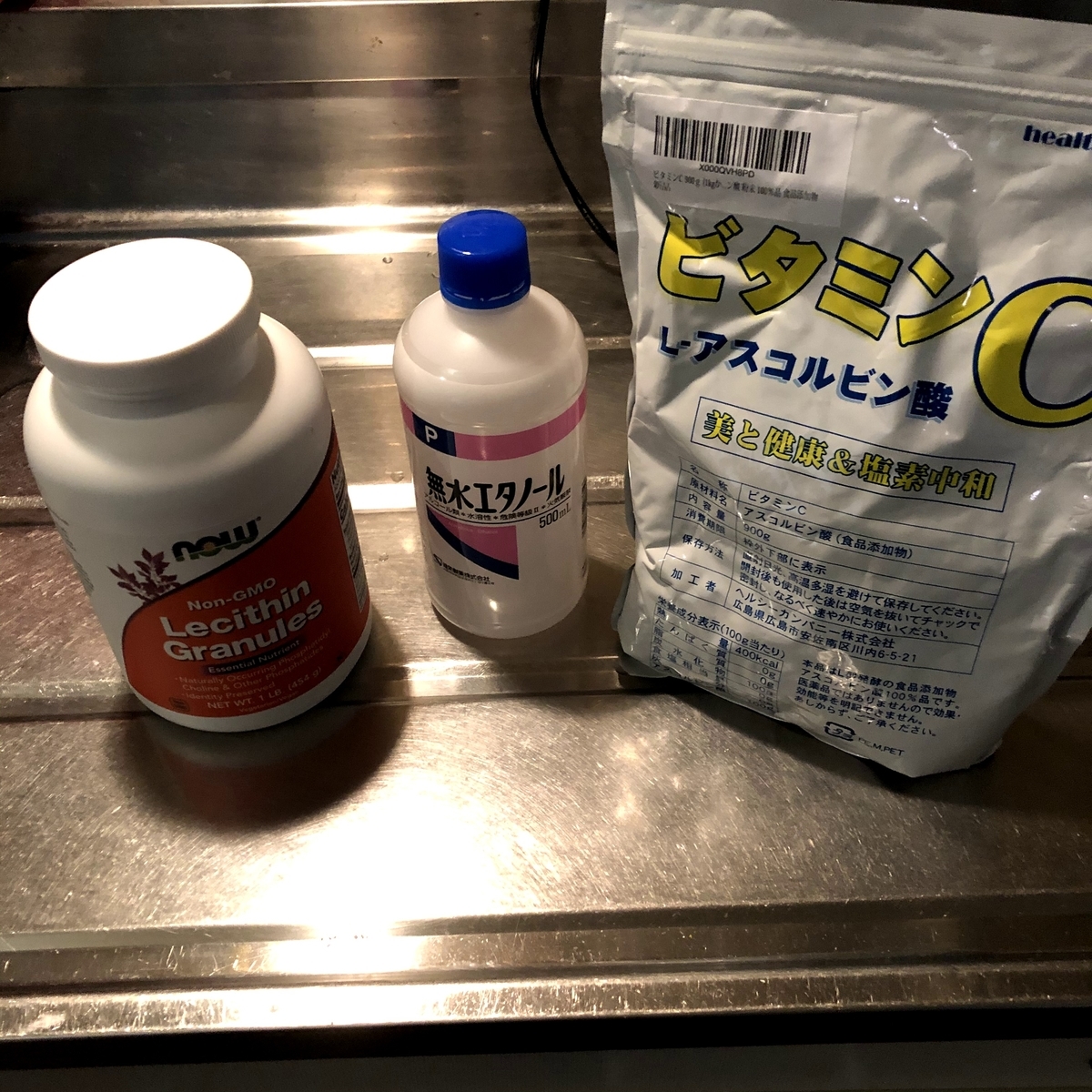
用意するもの
必須アイテム
・ビタミンC粉末
・顆粒レシチン
・無水エタノール
・温度計(100℃まではかれるものが望ましい)
・500mlビーカー
・キッチンスケール(0.1g単位ではかれるものが望ましい)
・水道水(契約しましょう)
あると便利なもの
・ガラス棒(混ぜれるものなら何でもOK、粘土が高いのでスプーンとかだと余計に付着してもったいない)
・任意のサイズのビーカー(キッチンスケールで量り取った材料を入れておくのに便利、容器なら何でもOK)
自作リポソームのレシピ
このレシピにおいては完成品の容量がおおよそ400mlになります。
準備
まず下準備としてそれぞれの材料を以下の重量分キッチンスケールなどで任意の容器に量り取っておいてください。
水 238.8g
エタノール 57.4g
ビタミンC 80.2g
レシチン 98.5g
※小数点以下は適当でいいです


①エタノール水溶液の作成

②ビタミンCを溶液に溶かす
①で生成したエタノール水溶液にビタミンCを溶かします。


右の写真では温度計は20℃を指しています。
常温では完全に溶解しないため湯煎するなどして40℃前後まで温めます。


右の写真では温度計が40℃を指しており、沈殿物がないためビタミンCが完全に溶解したことがわかります。
沈殿したビタミンC粉末が完全に溶解したことを確認したら次のステップに進みます。
③溶液とレシチンを混合
②で生成した溶液とレシチンをブレンダーに入れて4分ほど撹拌します。


②で生成した溶液にレシチンを十分に溶解させるために溶液の温度が40℃前後に温まっていることが理想的です。


少々わかりにくいですが上の写真はブレンダーで撹拌中の混合液です。


十分にレシチンと溶液が混ざったら一度冷蔵庫にいれて冷却します。
④撹拌と冷却を繰り返す
十分に冷えたらまたブレンダーで撹拌します。2~3分ほど撹拌したらまた冷蔵庫にいれて冷却します。できるだけ多くのレシチンをビタミンCと結合させるためにこのプロセスを2,3時間おきに合計で5,6回ほど繰り返します。
この時③の最初の撹拌をのぞいて液体の温度が32℃を超えないように注意してください(ブレンダーの撹拌による摩擦で温度が上昇します)。加熱しすぎるとカプセルが破壊されてしまいます。
このプロセスの完了によって溶液中のビタミンCの7割以上がレシチンと結合しカプセル化されます。
通常④のステップでリプライセルは完成ですが全てのビタミンCのカプセル化には至っていません。超音波洗浄機を用いることで100%カプセル化されたリプライセルを生成することが可能です。より完全なカプセル化を望まれる方はステップ⑤に進んでください。
超音波洗浄をお持ちでない方は⑤と⑥のステップはスキップし、ステップ⑦に進んでください。
⑤超音波照射による泡の除去
このステップでは混合液から泡を除去します。最後にもう一度ブレンダーで混合液を撹拌したあと、30度℃付近まで温まった混合液をビーカーにそそぎます。

このレシピにおいては500mlビーカーが丁度よいサイズになります。


また、上の写真のようにダンボールを適当な大きさに切って中央にビーカーの底部と同程度の半径の穴を作ることでより超音波の照射がしやすくなります。私は割り箸とセロテープを使って補強するなどしています。
混合液を入れたビーカーにラップをして超音波洗浄機の水槽に浸し30分ほど超音波を照射します。
超音波洗浄機付属の金網ネットは超音波出力が低下するため使わないことをおすすめします。

混合液の泡がビーカー上部にたまってきたらこのステップは完了です。冷蔵庫にいれてしばらく冷却します。
⑥断続的に超音波を照射
より完全なカプセル化のために断続的な超音波照射を行います。混合液の入ったビーカーを超音波洗浄機の水槽に浸し混合液の温度が32℃に達しないくらいの時間幅で超音波を照射します。1回につき20分ほどが目安と考えてください。
20分程度の超音波照射が完了したら冷蔵庫で1時間ほど冷却します。混合液が累積で1時間の超音波照射をうけるまでこのプロセスを繰り返します。1回につき20分の場合は3~4回このプロセスを繰り返すと良いでしょう。カプセル化が解除されないために温度は32℃を上回らないように注意してください。
⑦泡を捨てて完成
混合液の上部にたまった泡をスプーンなどですくって捨てた後、タッパーや瓶などの任意の保存容器に混合液をうつして完成です。

上の写真ではまだ泡が残っているのが確認できます。
保存は必ず冷蔵庫で行ってください。
このレシピにおいて作成された混合液には1mlあたり約200mgのカプセル化されたビタミンCが含まれています。これは大さじ一杯(15ml)あたり約3000mgのビタミンCが含まれていることになります。
用語解説
・レシチン
リン脂質の総称。ある時点まではリン脂質の一つであるホスファチジルコリンのみを指す用語であったが現在ではホスファチジルコリン、ホスファチジルセリン、ホスファチジルイノシトール、ホスファチジルエタノールアミン、及びその他多くのリン脂質全般を指す用語として定着している。レシチン - Wikipedia
2020/11/28:詳しい用語や器具の解説や製造途中の写真については追って加筆いたします。もう少々お待ち下さい。
2021/02/15:製造途中の写真を追加しました。
※本記事の情報において計算の参考となっている為替レートや販売価格は全て2020/11/28時点のものであり、有為転変の宇宙においてそれらは時間と共に常に変動します。
為替レートは2020/11/28 15:00時点における1ドル(USD)104.07円(JPY)のレートを参考にしています。
また、本記事の内容を元に自作したリプライセルを販売するなどして商業利用することはLivOn Laboratories社に対する特許侵害などの法的問題に発展する可能性があります。当ブログと当ブログの著者はそれらに係るあらゆる法的問題について一切責任を負いかねますのであらかじめご了承ください。
D City Rock feat. Debra Zeer
D City Rock feat. Debra Zeer
作詞 LISA
作曲 TeddyLoid
唄 TeddyLoid
My name is Panty, the crazy sexy blondie and I'm not dumb
(私の名前はパンティ、イカれたセクシーブロンドだけどバカじゃない)
I'm breaking the news, now boys! Us girls we're full-time horny too !
(ボーイズ、いいことを教えてやるよ!私ら女はいつだってエロい気分なんだ!)
Hey, check out that hot one, damn he's got a big one *** delicious
(ねえあのイケてるアイツ、あのビッグな***超おいしそう)
It's time to get dirty now, so will y'all excuse me read the air ! I'm busy !
(ヤりたくなってきたから悪いが失礼!空気読めよ!私は忙しいんだ!)
Garterbelt ! Again you've dialed D-CITY 2-900
(ガーターベルト!もう一度ダテンシティ2-900番に電話してみてよ!)
Pantsu line ! Copy that ! Clear the city !
(神のお告げよ!OK!街をキレイにしろとの指令だ!)
Anarchy ! Everyone wants to be me !
(アナーキー!みんな私になりたいんでしょ!)
Anarchy yeh ! Get it up so I can see !
(アナーキーにね!起きてよ!私にも見えるように!)
Anarchy ! Let it flow wild and free !
(アナーキー!本能と自由に身をまかせて!)
Anarchy yeh ! Ya'll ready for the gig ?!
(アナーキーにね!ライブの準備はできた!?)
By the way, my name is Stocking, I spill venom stinging eros
(そして私の名前はストッキング、あふれる毒舌でエロスに駆り立てるわ)
And I got sweet tooth for rollipops loli goth, I lick good !
These stripes will strip you down.
(このしましまがあなたたちを溶かしちゃうわ)
I got no mercy for chiralism
(チラ見せなんて許さないわ)
My fans they all love me cuz I kick it real tasty
(私がいい感じにぶっ飛ばせばいつもファンは私に夢中なの)
I get high being nasty !
(悪いことするのって最高!)
Garterbelt ! You keep slappin' my butt around
(ガーターベルト!あいつはいつも私のお尻を叩いてくるの)
Pantsu line ! Freaky girl comin' your way !
(神のお告げよ!イカしたガールの登場よ!)
Anarchy ! Everyone wants to be me !
(アナーキー!みんな私になりたいんでしょ!)
Anarchy yeah! Standing ovation please !
(アナーキーにね!スタンディングオベーションでよろしく!)
Anarchy ! Shake that thang wild and free !
(アナーキー!野生と自由で揺り動かして!)
Anarchy yeah ! Standing ready for the gig ?!
(アナーキにね!立って!ライブの準備はいい!?)
I know you know... those wings inside of you
(私は知ってるよ・・・あなたの中の2枚の翼)
I know you know... they do get naughty too
(わかってるよ・・・あいつらだっていけないことをしてるんだ)
Before I go...there's something I wanna say
(私が行ってしまう前に・・・言っておきたいことがあるの)
Your sleepy anarchy...Wake it up! Wake it up !
(あなたの眠れしアナーキーよ、立ち上がれ!羽ばたいて!)
Anarchy ! Everyone wants to be me !
(アナーキー!みんな私になりたいんでしょ!!)
Anarchy yeh ! Get it up so I can see !
(アナーキーにね!私にも見えるようにさ!!)
Anarchy ! Let it flow wild and free !
(アナーキー!本能と自由に身を任せてね!!)
Anarchy yeh ! Ya'll ready for the gig ?!
(アナーキーにさ!みんな準備はできてるんでしょ!!)
Although the future is a closed book
Anyone doesn't know what happens next moment.
Even Laplace's demon also does.
We don't have any choice except making the patterns as many as possible.
A current standard is no more than a current standard.
Certainly , the future is a closed book. But we can't know inside a closed book from what we can see now.
What we have to do as we prepare for indeterminate selection pressure is making and keeping diversity as much as possible.
I think it is nonsense to make evolution theory a basis for superior thought.
Because we can never judge what establishes the evolution finally , by the current standard , and in the current environment.
If we managed to carry out evolution based on the current standard and ideology , can that evolution keep up holding our superiority in the future , in the environment of indeterminate ?
This is the essential problem of fascism.
And then , also the reasonableness and benefits of the concept to be the center of the current human rights thought , like freedom , equality , love , and mutual recognition , is able to be explained by applying that theory.
With beyonding something religious , moral and ethical , this theory is quite concerned with the possibility that civilization and species can survive.
When we think about the ideal state of ideology , humanity and own way , we should always define prosperity and continuation of the civilization as a purpose.
In the future , we have to verify how each variation affects about life and death of the civilization systematically and logically at both micro and macro view.
In the future , we have to verify how each variation affects about life and death of the civilization systematically and logically at both micro and macro view.
次の瞬間何が起こるかは誰にもわかりません
ラプラスの悪魔にとってもそれは同じでしょう
唯一できることは最多のパターンを用意することです
現在の価値基準は現在の価値基準でしかありません
未来は未知であるが故に「未来」なのです
一寸先は闇ですが明るみから闇は探ることはできません
未知なる淘汰圧への最も堅固な対策は可能な限り多様化することです
ダーウィンの進化論を優勢思想の根拠とするのは非常にナンセンスと言えます
なにをもって進化とするかは現環境の現価値基準では判断しかねるからです
現時点の価値基準や思想を元に恣意的な進化を遂げたとして、それが未来の、未知の環境下での優位性を担保してくれるでしょうか
ファシズム体制の本質的な問題点はここにあるでしょう
また、平等、自由、博愛、相互承認といった今般の社会思想、人権思想の枢軸となる概念の合理性とメリットもこの理論を適用すれば説明がつきます
これは宗教や倫理、道徳上の問題に留まらず、実際に文明と種の存続可能性に大きく関係しています
私達は自分自身のあり方や思想や人道のあり方について考える際に、常に文明の繁栄と存続をそれらの目的として定義するべきだと思います
一つ一つの変化が文明の存亡にどのような影響を与えるか、それをミクロとマクロの両視点において体系的かつ論理的に検証することが求められる時代です
※文法や単語の間違いがあったらコメントなどで教えていただけると助かります